Advanced Technology
CAD/CAM
This technology is used in dentistry to fabricate crowns and bridges, dental implant abutments, dental implant crowns, and surgical guides. In our office it can be utilized to provide patients with a permanent crown in one appointment. These restorations are designed using computer software and milled (cut out) by machines. Some systems such as CEREC and E4D are complete systems that are smaller and able to be utilized in the dentist office. Larger systems such as BruxZir and Procera are typical only used in commercial dental laboratories. Our office currently uses CEREC and Procera Forte for most of our restorative needs.
Procera Forte is unique in that all restorations (mostly implant) are designed here in the office, however the milling units for Procera are housed in a central milling lab in New Jersey.
Cerec, as described above, refers to our 3D imaging system which can be used directly in the mouth or indirectly off a model of the patient’s mouth. The image is a digital 3D image which enables the computer to create a cyber 3 dimensional image of your teeth, and associated structures. The doctors can custom design your dental crown(s) and /or dental bridges for your teeth, dental implants, hybrids, etc. on the computer. This can be done quickly and as soon as a design is developed and ready for fabrication, the digital data is sent wirelessly directly to our in office milling machine. This machine will mill your dental crown or bridge within minutes. The restoration is then finished by one of us or our staff, and sintered in a firing oven to crystallize the ceramic. The appropriate staining or coloring is added and glazed. The result is a beautiful natural all ceramic crown with great strength and esthetic value, and it has been customized to blend and match your surrounding dentition in size, shape, position, surface texture and finally shade.
These crowns can easily be scheduled for the same appointment, all in one from preparation to final bonding, or, the patient can be released to go shopping, have lunch, etc. while we are completing the tasks Toward the end of the day the patient can return to complete the procedures. This is particularly attractive our patients who live out of town or out of country.
A further benefit of this technology is the fact that each and every crown or bridge is stored in the computer. Should there ever be an instance where a restoration is damaged , chipped, etc. in any way, the patient can call us in advance and we can fabricate a replacement from our archives before the patient arrives if it is appropriate.
Digital Imaging
Our office chooses carefully which and when radiographs are taken. There are many guidelines that we follow. Radiographs allow us to see everything we cannot see with our own eyes. Radiographs enable us to detect cavities in between your teeth, determine bone level, and analyze the health of your bone. We can also examine the roots and nerves of teeth, diagnose lesions such as cysts or tumors, as well as assess damage when trauma occurs. Dental radiographs are invaluable aids in diagnosing, treating, and maintaining dental health. Exposure time for dental radiographs is extremely minimal. Our office utilizes Digital Imaging Technologies within the office. With digital imaging, exposure time is about 50 percent less when compared to traditional radiographs. Digital imaging can also help us retrieve valuable diagnostic information. We may be able to see cavities better. Digital imaging allows us to store patient images, and enables us to quickly and easily transfer them to specialists or insurance companies.
Digital X-Rays:
Digital X-rays offer more precision since we view the image on a computer monitor, instead of holding up a 35mm film up to the light. Digital X-rays results in 1⁄6th the radiation exposure to you. Often times these may be utilized to provide a general idea if dental implants are possible.
Digital Cone Beam Computer Topography (CBCT):
 CBCT ScannerOur in office CBCT scanner allows us to generate a 3 dimensional rendering of a patients upper and lower jaw. When evaluating a patient for dental implants, this type of diagnostic information is essential when evaluating potential dental implant sites. Bone quality and quantity can be evaluated. A traditional 2 dimensional x-ray only allows for bone height evaluation but not bone width.
The viewing software allows us to evaluate and accurately measure bone height and width as well as accurately locate major nerves, maxillary sinuses, adjacent roots, nasal passages and irregular bone contours. This software also contains a library of different dental implant companies and all their sizes which can be “virtually” placed knowing precisely what size implants can be utilized and whether or not any bone grafting might be needed.
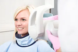
Intra-oral Camera
It is true. A picture is worth a thousand words. The intra-oral camera allows us to take up close detailed pictures of teeth showing decay, fractures, infection, and other pathologies. Dentists often lose touch with the fact that patients do not see what they see. Often times, when patients see these images, they are ready to make changes that might not be necessary. Education is essential in a doctor patient relationship. An educated patient is better able to make decisions they might otherwise need to depend on the doctor to make. In dentistry there are MANY “forks in the road”. Understanding the risk/reward allows the patient to choose what might be best for them at that time or long term. Almost all dental treatment is irreversible. This is often what influences the conservative nature of most dentists.
VELSCOPE
Velscope involves the use of a special light to help identify oral mucosal abnormalities. These abnormalities can be the early stages of cancer and might otherwise be difficult to see under normal light. It must be emphasized that Velscope does not provide a definitive diagnosis. Cancerous tissue can only be identified via tissue biopsy.
Laser Dentistry
Our office utilizes two different laser devices: soft and hard tissue.
Soft Tissue Laser:
This device is often used to remove excess gum tissue to improve aesthetics, provide hemostasis, or facilitate crown and bridge impressions.
Areas of dental care that benefit from laser technology:
- Cavity diagnosis and removal
- Curing, or hardening, bonding materials
- Whitening teeth
- Periodontal, or gum related, care
- Pediatric procedures
- Aphthous Ulcer treatment (canker sore)
- Frenectomy (tongue-tie release) without anesthesia or sutures
- Root canals and apicoectomies
- Crown lengthening, gingivectomy and other gum corrections
Dental lasers have been shown to be safe and effective for treating both children and adults.
Many patients, especially younger patients, are very familiar with the latest technology and are comfortable with the high tech practice. Computers and TV screens are their primary method of information processing. Dr. Bryhn Simmons utilizes intra-oral camera technology that helps enhance your understanding of your diagnosis. An intra-oral camera is a very small camera – in some cases, just a few millimeters long. An intra-oral camera allows our practice to view clear, precise images of your mouth, teeth and gums, in order for us to accurately make a diagnosis. With clear, defined, enlarged images, you see details that may be missed by standard mirror examinations. This can mean faster diagnosis with less chair-time for you! Intra oral cameras also enable our practice to save your images in our office computer to provide a permanent record of treatments. These images can be printed for you, other specialists, and your lab or insurance companies.